Vitamin K and Covid 19
COVID-19 and Vitamin K
- COVID-19 is a global pandemic, where some patients develop pulmonary infections and multiple micro-clots throughout the body.
- Vitamin K is required for both the anti-coagulation system to work, as well as for the better known coagulation system to work.
- The prevalence of micro-clots suggests that the anti-coagulation system has been compromised, raising questions about the sufficiency of vitamin K in those patients.
- Elastin is a key protein in connective tissue, such as in the lungs and veins.
- Matrix Gla protein is dependent on vitamin K to activate.
- Elastin needs MGP to prevent degradation and to maintain lung function.
- Insufficient amounts of vitamin K are linked to some prominent elements of COVID, including compromised lung function, and excessive clotting.
COVID-19 and Vitamin K
This page is devoted to presenting the emerging research on vitamin K and COVID as it is published.
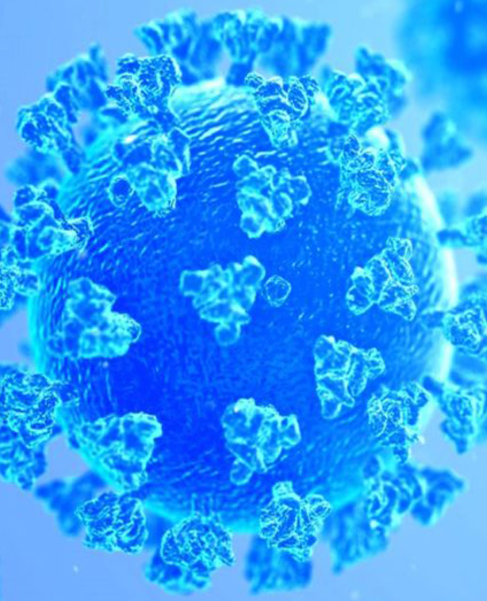
In 2019, a new coronavirus was identified as the cause of a disease outbreak that originated in China. Coronaviruses are named for the crown-like spikes on their surface.
The virus is now known as SARS-CoV-19. SARS is an acronym for Severe Acute Respiratory Syndrome. The acronym CoV refers to Corona Virus. The disease it causes is called COVID-19, with 19 referring to 2019 as the year in which it was identified. COVID is a significant global pandemic that the world is still in the midst of surviving.
COVID-19 was initially considered to be a respiratory virus and the majority of individuals who contract it have mild symptoms. A significant proportion do develop respiratory failure due to pneumonia (Zhou et al, 2020). However, COVID-19 symptoms may also manifest outside of the lungs, including generalized microvascular thrombosis, or clots, throughout body, as well as coagulopathy or impaired clotting, and excessive bleeding, which are associated with decreased survival [Cui et al, 2020; Lillicrap, 2020; Tank et al, 2020)]. The mechanisms leading from pulmonary infection to systemic coagulation dysfunction in Covid-19 have not yet been entirely understood.
Some of the research has focused on the coagulation/anti-coagulation system, in general, as well as elastin and matrix Gla protein. Elastin is a protein that makes up elastic connective tissue, and matrix Gla protein is a member of a family of vitamin K-dependent proteins.
Anti-Coagulation System
Based on the clotting cascade (see the Clotting webpage on this website), we know there are chemical elements and protein circulating through the bloodstream that are anticoagulant forces, that keep your blood liquid and flowing. These elements are proteins that are available when needed. Also key to anti-coagulation is the vascular system which the blood flows through, and which is lined with endothelial cells. Blood vessels consist of an impermeable inner layer of endothelial cells that are supported by elastic fibers and smooth muscle cells. Endothelial cells help regulate exchanges between the bloodstream and the surrounding tissues. The body has a potent system for anti-coagulation.
A naturally occurring anticoagulant protein produced in the liver and circulating through the body, is antithrombin (AT), which functions as a mild blood thinner. It acts like a police protein that prevents you from clotting too much. Antithrombin binds coagulation factors, inactivating their clotting potential (More et al, 1993; Liu & Rodgers, 1996; Broze, 1995; Stern et al, 1985; Shuman, 1986; Casu et al, 1981; Lindahl et al, 1980, Marcum & Rosenberg, 1984; Tollefsen & Pestka, 1985).
Also circulating are Protein C and Protein S, which are proteins in the blood that help regulate blood clot formation. Protein C works in conjunction with Protein S, and together they function as a natural anticoagulant system, mainly by inactivating clotting factors V and VIII, which are required for any clot generation. They work to prevent clots.
Endothelial cells secrete coagulation inhibitors on an ongoing basis (Stern et al, 1985; Fair et al, 1986), which locally help suppress clots (3-Fair et al, 1986). Some of these inhibitors include protein S, thrombomodulin, PA1-1, and TFPI. While Protein S circulates throughout the body, endothelial cells also synthesize and release Protein S, locally. Endothelial cells also regulate the activities of circulating blood cells, actively preventing platelets from gathering together, and they provide receptors for proteins in the blood that interfere with clot formation, like protein C.
These proteins in the anticoagulation system are vitamin K dependent. The three primary anticoagulation proteins, Protein C and Protein S and Protein Z, all depend on sufficient amounts of Vitamin K to be functional and, thus, active (Esmon et al 1987: Lobato-Mendizabal & Ruiz-Arguelles, 1990; Suttie, 1993; Grober et al, 2015). Ironically, while Vitamin K is well known for its role in helping blood to form clots, it is also very necessary to prevent clots from forming (Esmon et al, 1987: Lobato-Mendizabal & Ruiz-Arguelles, 1990; Grober, et al, 2015). Vitamin K is necessary to facilitate a potent system of anti-coagulation forces in the body, enhancing the effectiveness of the anti-coagulant system and keeping the blood fluid (Espana et al, 2005).
If the active or carboxylated forms of the proteins are low, due to insufficient vitamin K, then there can be increased coagulation in the blood vessels which can result in abnormal clotting, sometimes with devastating consequences (Dahlback, Villoutreix, 2005).
Keep in mind that the body operates on a triage theory, with the priority always being survival (McAnn, & Ames, 2009). This means that only after the needs of the clotting factors are met, will vitamin K be circulated throughout the body to other proteins outside of the liver. Hence, a vitamin K deficiency results in those proteins outside of the liver being more severely compromised than the hepatic vitamin K-dependent proteins found in the liver (Booth, Martini et al, 2003). This can paradoxically lead to enhanced thrombogenicity or enhanced clotting, in a state of low vitamin K (Nigwekar et al, 2018).
The anti-coagulant system is not triggered nor is it resolved by vitamin K. The anti-coagulant system functions regardless of the presence of vitamin K, however, its effectiveness in preventing clots is improved with sufficient amounts of vitamin K.
Coagulation System
The coagulation system depends on clotting factors, which are produced within the liver. The clotting factors are Factors II (thrombin), VII, IX, and X (Stenfloet al, 1934; Nelsestuen et al, 1974; Magnusson et al, 1974; Zytkovicz et al, 1975; Vermeer et al, 1978; Olson, 1984).
These four coagulation proteins need to be carboxylated or activated in order to become biologically functional, which requires adequate levels of vitamin K. Vitamin K enables those clotting factors to be activated and to clot the blood quickly (Askim, 2001; Bern, 2004; Theuwissen et al, 2012; Shearer & Newman, 2008).
Also circulating is procoagulant Factor II or prothrombin, which works with other clotting factors to form a clot.
Vitamin K is an essential “switch” in balancing coagulation and the anti-coagulation process (Espana et al, 2005). Vitamin K acts as a cofactor in the activation of proteins that that are involved in clotting and coagulation. On the other hand, vitamin K can also trigger key anticoagulants (Danziger, 2008).
Elastin Fibers
Elastin is a protein which forms the main element of elastic connective tissue. Elastin provides resilience, elasticity and deformability to dynamic tissues. Elastin fibers are essential matrix components in lungs and in blood vessels, and have high calcium affinity (Luo et al, 1997).
Elastic fiber degrades and is repaired on a continuous basis (Mithieux & Weiss, 2005) . Elastic fibers that are partially degraded become prone to mineralization (10). The balance between the degradation and the repair is delicate and of vital importance for cardiovascular and pulmonary health (Liu, et al, 2004).
The rate of elastic fibre degradation increases during ageing (Rabinovich et al, 2016). This age-related acceleration of elastolysis is enhanced in certain pulmonary conditions such as chronic obstructive pulmonary disease (COPD) and idiopathic pulmonary fibrosis (IPF) (Huang et al, 2016) .
MGP
Matrix Gla protein (MGP) is a well-known calcification inhibitor in soft tissues, such as arterial walls (Schurgers et al, 2013), and is expressed by the endothelial cells that line the vessel walls. Vascular calcification typically initiates in the elastin fibers of the arterial wall.
Besides preventing soft tissue calcification, MGP plays a critical role in protecting against pulmonary and vascular elastic fiber degradation and in the protection of elastic tissues against mineralization (Lee, et al, 2006). MGP is highly expressed in the lungs (Fraser & Price, 1988) and is important for pulmonary functioning (Price et al, 2001; Price, et al, 2009; Schurgers et al, 2013). This was demonstrated in MGP knockout mice, which developed severely mineralized as well as fragmented elastic fibers, when MGP was absent (Luo, Ducy et al, 1997)
Therefore, MGP’s role in the pulmonary compartment seems to be comparable with that in the vasculature and is crucial for the protection of elastic fibers against calcification. Impaired MGP activation could be associated with elastic fiber degradation. These processes are interrelated, and partially degraded elastic fibers are more prone to mineralization.
MGP is vitamin K-dependent, meaning it needs sufficient amounts of vitamin K to be active and functional.
Immune System
Vitamin K is also known to play an important role in modulating the immune system. Vitamin K is associated with an impaired production of inflammatory cytokines (Reddi et al, 1995; Ohsaki, et al, 2010; Chen et al, 2020). Interestingly, these are among the most important cytokines activated during SARS-CoV-2 infection (Wang et al, 2020), which contribute to a cytokine storm leading to ARDS in severe Covid patients (Ragab et al, 2020).
The alveolar-capillary membrane is the gas exchange surface of the lungs. It is the largest surface area within the lung and is the blood-air barrier in the gas exchanging region of the lungs. It prevents air bubbles from forming in the blood and it prevents blood from entering the alveoli. A hall mark of ARDS (Acute Respiratory Disease Syndrome) is the loss of the integrity of this membrane. Proteins C and S, which are activated by vitamin K play a protective role and help maintain the integrity of this membrane and modulate infection (Puig et al, 2013, Urawa, et al, 2016).
COVID-19
The early research has shown that vitamin K may be a key variable in the severity of COVID-19 outcomes via two pathways - a vitamin K deficiency in general and an increased demand for vitamin K due to the metabolism of elastin fiber from combatting the COVID-19 disease.
As discussed, vitamin K is necessary for both the anti-coagulation system and for the efficacy of the coagulation system when it is needed. A frequent outcome in severe COVID-19 cases are micro-clots throughout the vascular system and organs, indicating that the anti-coagulation system has been compromised. The first pathway is general vitamin K insufficiency, which would result in a deficient activation of endothelial protein S, a key protein in the anti-coagulation system (McCann & Ames, 2009). As discussed previously, if protein S works with protein C to inactivate clotting factors, and if protein S is less active due to insufficient vitamin K, then the stage is set for clotting to be unrestrained (Fair et al, 1986; Janssen et al, 2020).
This known pathway of vitamin K insufficiency that would trigger unrestrained clotting, aligns with the preliminary findings of the enhanced thrombogenicity in COVID-19 patients (Tang & Wang, 2020). Autopsies of people who died from severe COVID revealed bilateral, deep venous, leg thrombosis in all thromboembolic cases, and thrombosis of the prostatic venous plexus in the majority of men who died (Wichman et al, 2020)
In current research, Janssen (et al, 2020) proposed a mechanism of vitamin K depletion due to the onset of pneumonia, leading to a decrease in activated MGP and protein S, and resulting in pulmonary damage, elastic fiber damage, and clots. The pathologic lung involvement seen in COVID may consume M7, leading to the depletion (Mangge et al, 2022). Janssen recommends intervention trials to assess whether vitamin K administration plays a role in the prevention and treatment of severe COVID19. And unlike other treatment strategies, such as dexamethasone, vitamin K does not have any known unfavorable effects in those who do not use vitamin K antagonists. Its effectiveness could be rapidly evaluated and supplementing with vitamin K could be easily implemented if proven successful.
In a recent study, inactive vitamin Matrix Gla protein (dp-ucMGP) and prothrombin (PIVKA-II) (a clotting factor), were measured in 135 hospitalized COVID-19 patients. They found that inactive MGP was severely increased in the COVID patients, indicating vitamin K insufficiency compared to controls. In contrast, prothrombin procoagulant was preserved. The data suggested that the extra hepatic vitamin K (the amount circulating in the blood beyond the liver), was depleted, leading to an accelerated elastic fiber damage, as well as increased clotting in severe COVID-19 patients (Dofferhoff et al, 2020). They concluded that without sufficient vitamin K, it was not possible to activate enough MGP and the endothelial protein S to combat the symptoms of COVID (Fair et al, 1986). Furthermore low vitamin K status was associated with increased blood levels of desmosine, a biomarker of degradation of elastic fibres in the lung tissue (Piscaer et al, 2019).
A Danish study has found that low vitamin K status predicted mortality in patients hospitalized with COVID (Linneberg et al, 2020). They found that levels of vitamin K measures differed significantly between survivors and non-survivors.
Additionally, the consumption of clotting factors during thrombosis puts a further burden on vitamin K stores by increasing demand for activation of newly synthesized coagulation factors to replace used ones (Iba & Levy, 2020). The ongoing demand for vitamin K creates a progressive depletion of active endothelial protein S, which increasingly skews the balance towards coagulation.
The second pathway is the increased demand for vitamin K due to the metabolism of elastic fiber from the COVID-19 disease. Partially degraded and mineralized elastic are more vulnerable to further breakdown and calcification in comparison to intact fibers (Balsayga et al, 2004; Umeda et al, 2011). Upon SARS-CoV-2 infection, elastic fiber breakdown accelerates compared to degradation rates in age-matched controls (Dofferhof; Janssen et al, 2020). As MGP is protective of elastic fibers, there is an increased need for MGP synthesis to maintain and protect elastic fibers (Luo et al, 1997; Parker et al, 2009), further draining vitamin K stores and leading to higher levels of dp-ucMGP or uncarboxylated, inactive matrix Gla protein. There appears to be interrelationships between vitamin K shortage, insufficient MGP carboxylation and elastic fiber pathologies in COVID-19 (Liux et al, 2004; Mithieux & Wiess, 2005). Vitamin K is considered to promote vascular health and reduces the risk of developing lung scarring in COVID-19.
It has also been observed that many COVID-19-infected individuals who suffer from certain premorbid conditions, such as hypertension, diabetes, cardiovascular disease, atherosclerosis, and obesity, have an increased risk of complicated disease course (Huang et al, 2020). Although these conditions are also associated with poor outcomes due to other infectious diseases, these disorders are already associated with elastic fiber pathologies, as well as vitamin K insufficiency. Partially degraded and mineralized elastic fibers are quite prevalent in diabetic, hypertensive, renal and cardiovascular patients (Balsayga et al, 2004; Umeda et al, 2011). Matrix Gla protein is necessary for the inhibition of elastic fiber mineralization and degradation. The research has already demonstrated a correlation between matrix Gla protein, and elastic fiber degradation and arterial calcification (Liux et al, 2004; Mithieux & Weiss, 2005). So, if a person has a comorbidity such as hypertension or diabetes, their system is vulnerable to elastin fiber degradation and would need extra vitamin K to activate MGP so as to defend and maintain their current health status, once infected with COVID-19.
It is also interesting that many comorbid conditions, which worsen COVID-19 clinical outcomes, are associated with a compromised level of MK-7 (Kang & Jung, 2020). An MK-7 deficiency is certainly linked to vascular calcification, as it reduces the concentration of active MGP to below that which is required for the inhibition of elastic fiber mineralization. Therefore, MK-7 deficiency can be a risk factor for increasing the severity of the COVID-19 disease, and SARS-CoV-2 infected patients with comorbid conditions do tend to develop acute manifestations. Evidence suggests that similar processes also occur in the lungs. Calcification of elastic fibers makes them more susceptible to degradation, with subsequent disease complications. A clinical trial to assess the change in the MK-7 status of individuals before and after supplementation would be required to determine the effect of MK-7 on SARS-CoV-2 pneumonia. An Austrian study looked at vitamin K1, MK4, MK7 and vitamin K epoxide levels in healthy controls, and patients with non COVID-19 pneumonia, and hospitalized COVID-19 patients. They found that the COVID-19 patients had significantly lower MK7 levels when compared to the other two groups, as well as significantly decreased vitamin K1 and increased MK4 levels. MK4 was lowest in the covid patients, suggesting is was a fundamental part of the inflammatory and oxidative stress factors (Mangge et al, 2022).
Janssen (et al, 2020) proposes a series of sequential pathological steps that may occur in response to COVID-19 infection, which are responsible for upregulation of MGP expression, and the extrahepatic vitamin K depletion, leading to pulmonary damage and thrombosis in Covid-19.
It also appears that a potential relationship exists between MK-7 deficiency, lung tissue injury, and comorbidities in COVID-19 patients (Berenjian, 2020).
Vitamin D has been correlated with a heavily reduced fatality rate for COVID-19 patients. A frequent argument against supplementation of vitamin D2 is that high intakes could lead to hypercalcemia, which is the buildup of calcium in the blood (Orme et al, 2016). However, the hypercalcemia is typically the result of a vitamin K deficiency (Vermeer & Theuwissen, 2011). When vitamin D2 is supplemented with vitamin K, there are no long term health risks from vitamin D, and patients with COVID would benefit from the immunoregulatory effects and the increased chances of survival (Maghbooli et al, 2020; Karonova et al, 2020).
In conclusion, the potential role of vitamin K supplementation to prevent the development and progression of severe Covid-19 remains largely unexplored, but very promising (Kudelko et al, 2021; Samad et al, 2021). The diverse and distinct roles of vitamin K in modulating blood clotting, elastin degradation, immunomodulation, and managing vascular health, together with the low toxicity of vitamin K in humans, makes vitamin K an attractive remedy (Kudelko et al, 2021).
There is a need for further experimental evidence to link vitamin K deficiency with the pathology of Covid-19 and to determine whether vitamin K supplementation has a place in treatment protocols. Clinical trials are essential to evaluate whether MK-7 administration improves the health outcome or prognosis of patients with COVID-19. Thankfully, these clinical trials have begun, one being with Kappa Bioscience entering into a research collaboration with US based hospitals to explore the role of vitamin K2 status and COVID severity in hospitalized patients.
One recent trial studied hospitalized patients in the Netherlands. They found that levels of vitamin K were more significant than vitamin D, relative to patient outcomes. Vitamin K status was heavily associated with COVID inflammation, as measured by Interleukin-6. And IL-6 was significantly associated with elastic fiber degradation, a key marker of lung damage. Lowering Interleukin-6 is an important treatment goal for patients with severe COVID. Patients with good outcomes had higher levels of vitamin K (Visser et al, 2022).
Certainly, the impact of the current crisis warrants a thorough evaluation of the therapeutic potential of vitamin K in Covid-19 pathogenesis for two key reasons. Unlike other treatment strategies currently under development for Covid-19 such as dexamethasone, vitamin K does not have any known unfavorable effects in those who do not use vitamin K antagonists. Furthermore, it is relatively simple to manufacture, contrary to other therapies. Taken together this means that effectiveness can be rapidly and cheaply evaluated in clinical trials and easily implemented if proven successful.
References
1970
Stenflo J, Fernlund P, Egan W, Roepstorff P. Vitamin K dependent modifications of glutamic acid residues in prothrombin. Proceedings of the National Academy of Sciences of the United States of America. 1947;71(7):2730.
Nelsestuen GL, Zytkovicz TH, Howard JB. The mode of action of vitamin K. Identification of gamma-carboxyglutamic acid as a component of prothrombin. J Biol Chem. 1974 Oct 10;249(19):6347-50.
Magnusson SL, Slottrup-Jensen TF, Petersen HR, Morris HR, Dell A. Primary structure of the vitamin K-dependent part of prothrombin. FEBS Lett. 1974;44:189-93.
Zytkovicz TH, Nelsestuen GL. [3h] diborane reduction of vitamin K-dependent calcium-binding proteins. Identification of a unique amino acid. J Biol Chem. 1975 Apr 25;250(8):2968-2972.
Vermeer C, Govers-Riemslag JW, Soute BA, Lindhout MJ, Kop J, Hemker HC. The role of blood clotting factor V in the conversion of prothrombin and a decarboxy prothrombin into thrombin. Biochim Biophys Acta. 1978 Feb 1;538(3):521-33.
1980
Olson. RE. The function and metabolism of vitamin K. Ann Rev Nutr. 1984;4:281-337.
Fair DS, Marlar RA & Levin EG. Human endothelial cells synthesize protein S. Blood. 1986;67:1168-1171.
Edothelial cells, via the production and expression of protein S, may play a significant regulatory role in the initiation, propogation and suppression of hemostasis and thrombosis.
Stern D, Brett J, Harris K, et al. Participation of endothelial cells in the protein C protein S anticoagulant pathway: the synthesis and release of protein S. J Cell Biol. 1986;102:1971-1978.
Alperin JB. Coagulopathy caused by vitamin K deficiency in critically ill, hospitalized patients. JAMA. 1987;258:1916-1919.
Fraser J.D., Price P.A. Lung, heart, and kidney express high levels of mRNA for the vitamin K-dependent matrix Gla protein. Implications for the possible functions of matrix Gla protein and for the tissue distribution of the gamma-carboxylase. J. Biol. Chem. 1988;263:11033–11036.
1990
Luo G, Ducy P, McKee MD, et al. Spontaneous calcification of arteries and cartilage in mice lacking matrix GLA protein. Nature. 1997;386:78-81.
Reddi K, Henderson B, Meghji S, Wilson M, Poole S, Hopper C, et al. Interleukin 6 production by lipopolysaccharide-stimulated human fibroblasts is potently inhibited by naphthoquinone (vitamin K) compounds. Cytokine. 1995;7:287-290.
2000
Askim M. Vitamin K in the Norwegian diet and osteoporosis. Tidsskr Nor Laegeforen. 2001 Sep 20;121(22):2614-6.
There is a discussion in the literature of whether the allowances should be considerably higher (375 micrograms K1/day). Deep green vegetables and soybean oil are the best sources of vitamin K1, while cheese gives some K2. On the basis of this knowledge about the importance of vitamin K and osteoporosis, an intervention test should be done with respect to the high incidence of osteoporosis in Norway. Analysis of Norwegian foods for vitamin K1 and K2 is needed.
Price PA, Buckley JR & Williamson MK. The amino bisphosphonate ibandronate prevents vitamin D toxicity and inhibits vitamin D-induced calcification of arteries, cartilage, lungs and kidneys in rats. J Nutr. 2001;131:2910-2915.
Basalyga, DM, Simionescu, DT, Xiong, W, et al. Elastin degradation and calcification in an abdominal aorta injury model: role of matrix metalloproteinases. Circulation. 2004;110:3480–3487.
Fair DS, Marlar RA, Levin EG. Human endothelial cells synthesize protein S. Blood 1986; 67(4): 1168-71.
Booth SL, Martini L, Peterson JW, Saltzman E, Dallal GE, Wood RJ. Dietary phylloquinone depletion and repletion in older women. J Nutr 2003; 133(8): 2565-9.
Bern M. Observations on possible effects of daily vitamin K replacement, especially upon warfarin therapy. JPEN. 2004;28:388-98.
Liu X, Zhao Y, Gao J, et al. Elastic fiber homeostasis requires lysyl oxidase-like 1 protein. Nat Genet. 2004;36:178-182.
Espana F, Medina P, Navarro S, Zorio E, Estelles A, Zanar J. The multifunctional protein C system. Curr Med cham Cariovas Hematol Agents. 2005;3:119-31.
Mithieux SM & Weiss AS. (2005) Elastin. Adv Protein Chem. 2005;70:437-461.
Elastin is a key extracellular matrix protein that is critical to the elasticity and resilience of many vertebrate tissues including large arteries, lung, ligament, tendon, skin, and elastic cartilage. Knowledge of the major stages in elastin assembly has facilitated the construction of in vitro models of elastogenesis, leading to the identification of precise molecular regions that are critical to elastin-based protein interactions.
Lee J.S., Basalyga D.M., Simionescu A., Isenburg J.C., Simionescu D.T., Vyavahare N.R. Elastin calcification in the rat subdermal model is accompanied by up-regulation of degradative and osteogenic cellular responses. Am. J. Pathol. 2006;168:490–498.
Danziger J. Vitamin K-dependent proteins, warfarin and vascular calcification. Clin J Am Soc Nephrol. 2008;3:1504-10.
Shearer MJ, Newman P. Metabolism and cell biology of vitamin K. Thromb Haemost. 2008Oct;100(4):530-47.
Burstyn-Cohen T, Heeb MJ & Lemke G. (2009) Lack of protein S in mice causes embryonic lethal coagulopathy and vascular dysgenesis. J Clin Invest. 2009;119:2942-2953.
Protein S (ProS) is a blood anticoagulant encoded by the Pros1 gene, and ProS deficiencies are associated with venous thrombosis, stroke, and autoimmunity.
McCann, JC & Ames, BN. Vitamin K, an example of triage theory: is micronutrient inadequacy linked to diseases of aging? Am J Clin Nutr. 2009;90:889–907.
The triage theory posits that some functions of micronutrients (the approximately 40 essential vitamins, minerals, fatty acids, and amino acids) are restricted during shortage and that functions required for short-term survival take precedence over those that are less essential. Insidious changes accumulate as a consequence of restriction, which increases the risk of diseases of aging. A triage perspective reinforces recommendations of some experts that much of the population and warfarin/coumadin patients may not receive sufficient vitamin K for optimal function of Vitamin K-Dependent proteins that are important to maintain long-term health.
Parker, BD, Ix, JH, Cranenburg, EC, et al. Association of kidney function and uncarboxylated matrix Gla protein: data from the Heart and Soul Study. Nephrol Dial Transplant. 2009;24:2095–2101.
Price PA, Toroian D, Lim JE. Mineralization by inhibitor exclusion: the calcification of collagen with fetuin. J Biol Chem. 2009;284:17092-101.
2010
Ohsaki Y, Shirakaw H, Miura A, Giriwono PE, Sato S,Ohashi A, et al. Vitamin K suppresses the lipopolyssacharide-induced expression of inflammatory cytokines in cultured macrophage-like cells via the inhibition of the activation of nuclear factor κB through the repression of IKKα/β phosphorylation. J Nutr. Biochem. 2010;21:1120-26.
We previously found that vitamin K suppresses the inflammatory reaction induced by lipopolysaccharide (LPS) in rats and human macrophage-like THP-1 cells. In this study, we further investigated the mechanism underlying the anti-inflammatory effect of vitamin K by using cultures of LPS-treated human- and mouse-derived cells. All the vitamin K analogues analyzed in our study exhibited varied levels of anti-inflammatory activity. These results clearly show that the anti-inflammatory activity of vitamin K is mediated via the inactivation of the NFκB signaling pathway.
Umeda, H, Aikawa, M & Libby, P. Liberation of desmosine and isodesmosine as amino acids from insoluble elastin by elastolytic proteases. Biochem Biophys Res Commun. 2011;411:281–286.
Vermeer C, Theuwissen E. Vitamin K, osteoporosis and degenerative diseases of ageing. Menopause Int. 2011;17:19-23.
Theuwissen E, Smit E, Vermeer C. The role of vitamin K in soft-tissue calcification. Advances in Nutr. 2012;3(2):166-173.
MGP is synthesized by vascular smooth muscle cells and is the most important inhibitor of arterial mineralization currently known. Carboxylation of the most essential Gla proteins take place in the liver, and that of the less essential Gla proteins take place in the extrahepatic tissues. A transport system has evolved to ensure preferential distribution of dietary vitamin K to the liver, when supplies are short. Research has shown that during a three year supplementation period, those who received vitamin K maintained their vascular elasticity, compared to a control group with a parallel 12% loss of elasticity.
Puig F, Fuster G, Adda M, Blanch L, Farre R, Navajas D, et al. Barrier-protective effects of activated protein C in human alveolar epithelial cells. PloS ONE. 2013;8:e56965.
Schurgers LJ, Uitto J & Reutelingsperger CP. Vitamin K-dependent carboxylation of matrix Gla-protein: a crucial switch to control ectopic mineralization. Trends Mol Med. 2013;19:217-226.
This review focuses on molecular mechanisms of vascular mineralization involving MGP and discusses the potential for treatments and biomarkers to monitor patients at risk for vascular mineralization.
Huang JT, Bolton CE, Miller BE, et al. Age-dependent elastin degradation is enhanced in chronic obstructive pulmonary disease. Eur Respir J. 2016;48:1215-1218.
Orme RP, Middleditch C, Waite L, Fricker RA. Chapter 11 – The role of vitamin D2 in the development and neuroprotection of midbrain Dopamine neurons. In: Litwack G, editor. vitamins & hormones, vitamin D hormone. Cambridge, Academic Press; 2016, p. 273-297.
Rabinovich RA, Miller BE, Wrobel K, et al. Circulating desmosine levels do not predict emphysema progression but are associated with cardiovascular risk and mortality in COPD. Eur Respir J. 2016;47:1365-1373.
Urawa M, Kobayashi T, D’Allesandro-Gabbazo CN, Fujimoto H, Toda M, Roeen Z, et al. Protein S is protective in pulmonary fibrosis. J Thromb Haemost. 2016;14:1588-99.
Janssen, R & Vermeer, C. Vitamin K deficit and elastolysis theory in pulmonary elasto-degenerative diseases. Med Hypotheses. 2017;108:38–41.
Elastin is a unique protein providing deformability and resilience to dynamic tissues, such as arteries and lungs. It is an absolute basic requirement for circulation and respiration. Elastin calcification and elastin degradation are two pathological processes that impair elastin's functioning. Matrix Gla Protein (MGP) is probably the most potent natural inhibitor of elastin calcification and requires vitamin K for its activation. The 'Vitamin K deficit and elastolysis theory' posits that elastin degradation causes a rise in the vitamin K deficit and implies that vitamin K supplementation could be preventing elastin degradation. If this hypothesis holds true and is universally found in every state and condition, it will have an unprecedented impact on the management of every single pulmonary disease characterized by accelerated elastin degradation, such as alpha-1 antitrypsin deficiency, bronchiectasis, COPD and cystic fibrosis.
Lindholt JS, Frandsen NE, Fredgart MH, Ovrehus KA, Dahl JS, et al. Effects of menaquinone-7 supplementation in patients with aortic valve calcification: study protocol for a randomized controlled trial. BMJ Open. 2018;8:3022019.
In this study, 400 men, aged 65-74 with aortic valve calcification will receive MK-7 (720 µg/day) supplemented by the recommended daily dose of vitamin D (25 µg/day) or placebo treatment for two years. Calcification will be monitored with CT scans.
Nigwekar SU, Thadhani R, Brandenburg VM. Calciphylaxis. N Engl J Med 2018; 378(18):1704-14.
Chen, HG, Sheng, LT, Zhang, YB, et al. Association of vitamin K with cardiovascular events and all-cause mortality: a systematic review and meta-analysis. Eur J Nutr. 2019;58;2191–2205.
Piscaer I, van den Ouweland JMW, Vermeersch K, et al. Low Vitamin K Status Is Associated with Increased Elastin Degradation in Chronic Obstructive Pulmonary Disease. J Clin Med. 2019;8(8). doi:10.3390/jcm8081116.
Elastin degradation is accelerated in chronic obstructive pulmonary disease (COPD) and is partially regulated by Matrix Gla Protein (MGP), via a vitamin K-dependent pathway. The aim of this study was to assess vitamin K status in COPD as well as associations between vitamin K status, elastin degradation, lung function parameters and mortality. A total of 192 COPD patients and 186 age-matched controls were included. In addition to this, 290 COPD patients from a second independent longitudinal cohort were also included. They found that reduced vitamin K status was found in COPD patients compared to smoking controls (p < 0.0005) and controls who had never smoked (p = 0.001). Vitamin K status was inversely associated with desmosine. Our results therefore suggest a potential role of vitamin K in COPD pathogenesis.
2020
Anastasi E, Lalongo C, Labriola R, Ferraguti G, Lucarelli M, Angeloni A. Vitamin K deficiency and covid-19. Scandinavian J of Clin and Lab Invest. 2020;DOI: 10.1080/00365513.2020.1805122
This study investigated the diagnostic role of vitamin K deficiency in the context of covid19 patients in Italy. They measured the serum level of PIVKA-II in a larger cohort of 62 covid19 patients and evaluated the correlation between IL-6 levels and vitamin K status. Vitamin K deficiency was observed in 41 patients with almost twice the incidence in males than females (72.3% vs 36.8% respectively). The prevalence of subjects with both IL-t and PIVKA-II above their respective cut off limits was significantly associated with the gender, males. They proposed two observations. The outcome of COVID for patients in the ICUis more severe in men than women, which may be related to the great IL-6 and lower vitamin K level in the male gender. And they postulate that the vitamin K deficiency arising during early phases of the COVID19 infection may contribute to the activation of the TH2 storm with increased production of IL-6. The Results highlight the role of vitamin K as a possible modifiable risk factor for a more severe evolution of covid19 in infected patients with clinicals symptoms.
Berenijan A. How menaquinone-7 deficiency influences mortality and morbidity in COVID-19 patients. Biocatal Agric Biotechnol. 2020 Oct; 29:101792.
Vitamin K is known for its role as a coagulation cofactor, and it naturally exists in two forms, vitamin K1 (phylloquinone) and vitamin K2 (menaquinone). Both vitamin subtypes act as a necessary cofactor for the carboxylation of vitamin K dependent proteins (Gla proteins). However, the two distinct forms of vitamin K are vastly different with respect to their chemical structure and pharmacokinetics that affect metabolism, bioavailability, and impact on health outcomes. The liver uses phylloquinone to activate coagulation factors, while menaquinone plays a more dominant role in the activation of extrahepatic vitamin K-dependent proteins, such as the matrix Gla protein (MGP). Therefore, phylloquinone primarily participates in blood clotting, and menaquinone is involved in important metabolic processes, such as bone mineralization and soft tissue decalcification. Therefore, this work aims to discuss how MK-7 can support the prevention and treatment of the disease during the COVID-19 pandemic.
Chen G, Wu, D. Guo W, Cao Y, Huang D, Wang et al. Clinical and immunological features of severe and moderate coronavirus disease. J Clin Investig. 2020;130:2620-29.
Cui S, Chen S, Li X, Liu S, Wang F. Prevalence of venous thromboembolism in patients with severe novel coronavirus pneumonia. J Thromb Haemost. 2020 Jun;18(6):1421-24.
Severe novel coronavirus pneumonia (NCP) patients have abnormal blood coagulation function, but their venous thromboembolism (VTE) prevalence is still rarely mentioned. 81 severe COVID patients in the ICU in Wuhan, China were studied. 25% (20) had venous thrombosis (clots), of which 8 died. The incidence of venous thrombosis seems to be related to a poor prognosis. An intervention trial is now needed to assess whether vitamin K administration improves outcome in patients with COVID-19 by increasing pulmonary MGP and endothelial protein S activation.
Dofferhoff ASM, Piscaer I, Schurgers, LJ, Visser MPJ, van den Ouweland JMW, de Jong PA, et al. Reduced vitamin K status as a potentially modifiable risk factor of severe COVID-19. Clin Infec Diseases. 2020 Aug 27; ciaa1258, https://doi.org/10.1093/cid/ciaa1258
Most patients infected with covid19 have mild symptoms, but a significant proportion develop respiratory failure. In severe cases, coagulopathy and thromboembolism are prevalent and related to decreased survival. Coagulation is an intricate balance between clot promoting and dissolving processes in which vitamin K plays a well-known role. We hypothesized that vitamin K status is reduced in patients with severe COVID-19. Vitamin K status was assessed by measuring desphospho-uncarboxylated matrix Gla protein (dp-ucMGP; inversely related to vitamin K status) and the rate of elastin degradation by measuring desmosine. We included 123 patients who were admitted with COVID-19 and 184 controls. These data suggest a mechanism of pneumonia-induced extrahepatic vitamin K depletion leading to accelerated elastic fiber damage and thrombosis in severe COVID-19 due to impaired activation of MGP and endothelial protein S, respectively. Measures of low vitamin K levels were significantly higher in COVID-19 patients with unfavorable outcome compared to those with less severe disease, and that low vitamin K status seems to be associated with accelerated elastin degradation. An intervention trial is now needed to assess whether vitamin K administration improves outcome in patients with COVID-19.
Huang C, Wang Y, Li X, et al. Clinical features of patients infected with 2019 novel coronavirus in Wuhan, China. Lancet. 2020;395:497-506.The 2019-nCoV infection caused clusters of severe respiratory illness similar to severe acute respiratory syndrome coronavirus and was associated with ICU admission and high mortality. Of 41 admitted hospital patients, testing positive for COVID, 73% were men, 32% had underlying diseases, median age was 49, 98% had a fever, 76% had a cough, 44% had fatigue, among other symptoms. All 41 patients had pneumonia. 32% were admitted to the ICU and 15% died.
Janssen R, Visser MPJ, Dofferhoff, ASM, Vermeer C, Janssens W, Walk J. Vitamin K metabolism as the potential missing link between lung damage and thromboembolism in Covid-19. Br J Nutr. 2020 Oct; 1-25.
Coronavirus disease 2019 (Covid-19), caused by SARS-CoV-2, exerts far-reaching effects on public health and socioeconomic welfare. The majority of infected individuals have mild to moderate symptoms but a significant proportion develops respiratory failure due to pneumonia. Thrombosis is another frequent manifestation of Covid-19 that contributes to poor outcomes. Vitamin K plays a crucial role in activation of both pro- and anticlotting factors in the liver, and the activation of extrahepatically synthesized protein S which seems to be important in local thrombosis prevention. However, the role of vitamin K extends beyond coagulation. Matrix Gla protein (MGP) is a vitamin K-dependent inhibitor of soft tissue calcification and elastic fibre degradation. Severe extrahepatic vitamin K insufficiency was recently demonstrated in Covid-19 patients, with high inactive MGP levels correlating with elastic fibre degradation rates. This suggests that insufficient vitamin K-dependent MGP activation leaves elastic fibres unprotected against SARS-CoV-2 induced proteolysis. In contrast to MGP, Covid-19 patients have normal levels of activated factor II, in line with previous observations that vitamin K is preferentially transported to the liver for activation of procoagulant factors. We therefore expect that vitamin K-dependent endothelial protein S activation is also compromised, which would be compatible with enhanced thrombogenicity. Taking these data together, we propose a mechanism of pneumonia-induced vitamin K depletion, leading to a decrease in activated MGP and protein S, aggravating pulmonary damage and coagulopathy, respectively. Intervention trials should be conducted to assess whether vitamin K administration plays a role in prevention and treatment of severe Covid-19.
Iba, T & Levy, JH. Sepsis-induced coagulopathy and disseminated intravascular coagulation. Anesthesiology. 2020;132:1238–1245.
Karanova TL, Andreeva AT, Vashukova MA. Serum 25 (oH) level in patients with CoVID-19. J Infectology. 2020;12(3): https://doi.org/10.22625/2072-6732-2020-12-3-21-27.
Vitamin D deficiency is considered as a risk factor for the incidence and severity of CoVid-19 infection. This study looked at 80 patients aged 18 to 94 years with severe and moderate CoVID. IT was found that in patients with a severe course, the level of vitamin D was significantly lower and more common, when compared to patients who had a moderate course of COVID. They concluded that vitamin D deficiency and obesity increased the risk of severe course and death of coronavirus infection.
Kang SJ, Jung SI. Age-Related morbidity and mortality among patients with COVID-19. Infec Chemother. 2020 Jun;52(2):154-164.
On March 11, 2020, the World Health Organization declared coronavirus disease (COVID-19), caused by the novel coronavirus severe acute respiratory syndrome coronavirus 2 (SARS-CoV-2), a pandemic. During the COVID-19 pandemic, an age-associated vulnerability in the burden of disease has been uncovered. Understanding the spectrum of illness and the pathogenic mechanism of the disease in a vulnerable population is critical, especially during the pandemic. Herein, we reviewed published COVID-19 epidemiology data from several countries to identify any consistent trends in the relationship between age and COVID-19-associated morbidity or mortality. We also reviewed the literature for studies explaining the difference in the host response to SARS-CoV-2 infection according to age. The insights from these data will be useful in determining the treatment policies and preventive measures of COVID-19.
Lillicrap D. Disseminated intravascular coagulation in patients with 2019-nCoV pneumonia. J Thromb Haemost. 2020 Apr;18(4):786-87.
Linneberg A, Kampmann FB, Israelsen SB, Andersen LR, Jørgensen HL, Sandholt H, et al. Low vitamin K status predicts mortality in a cohort of 138 hospitalized patients with COVID-19. Preprint: medRxiv 2020. 12.21.20238613;doi:https://doi.org/10.1101/2020.12.21.20248613.
This study tested the hypothesis that low vitamin K status is common in patients hospitalized with COVID-19 compared to population controls; and that low vitamin K status predicts mortality in COVID-19 patients. They measured blood levels of dp-uc Matrix Gla Protein which reflects functional vitamin K status in peripheral tissue. They found that low vitamin K status was associated with higher mortality risk, and also predicted mortality in patients. This data supports a potential role of vitamin K in COVID-19.
Maghbooli Z, Ebrahimi M, Shirvani A, Nasiri M, Pasoki M, Kafan S. Social Science Research Network; Rochester, NY: 2020. Vitamin D Sufficiency Reduced Risk for Morbidity and Mortality in COVID-19 Patients. PloS One. 2020;
Ragab D, Salah Eldin H, Taeimah M, Khattab R, Salem R. The COVID-19 cytokine storm; What we know so far. Front Immunol. 2020;11:1446.
Tang N, Li D, Wang X, et al. (2020) Abnormal coagulation parameters are associated with poor prognosis in patients with novel coronavirus pneumonia. J Thromb Haemost. 2020;18:844- 847.
Conventional coagulation results and outcomes of 183 consecutive patients with confirmed NCP in Tongji hospital were retrospectively analyzed. The overall mortality was 11.5%, the non-survivors revealed significantly higher D-dimer and fibrin degradation product (FDP) levels, longer prothrombin time and activated partial thromboplastin time compared to survivors on admission (P < .05); 71.4% of non-survivors and 0.6% survivors met the criteria of disseminated intravascular coagulation during their hospital stay.
Tutusaus A, Mari M, Ortiz-Perez JT, Nicolaes GAF, Morales A, Garcia de Frutos P. Role of vitamin K-dependent factors Protein S and GAS6 and TAM receptors in SARS-CoV-2 infection and COVID-19-Associates immunothrombosis. Cells. 2020;9:2186.
The vitamin K-dependent factors protein S (PROS1) and growth-arrest-specific gene 6 (GAS6) and their tyrosine kinase receptors TYRO3, AXL, and MERTK, the TAM subfamily of receptor tyrosine kinases (RTK), are key regulators of inflammation and vascular response to damage. TAM signaling, which has largely studied in the immune system and in cancer, has been involved in coagulation-related pathologies. Because of these established biological functions, the GAS6-PROS1/TAM system is postulated to play an important role in SARS-CoV-2 infection and progression complications. In the context of COVID-19, the role of TAMs could provide new clues in virus-host interplay with important consequences in the way that we understand this pathology. From the viral mimicry used by SARS-CoV-2 to infect cells, to the immunothrombosis that is associated with respiratory failure in COVID-19 patients, TAM signaling seems to be involved at different stages of the disease. TAM targeting is becoming an interesting biomedical strategy, which is useful for COVID-19 treatment now, but also for other viral and inflammatory diseases in the future.
Wang J, Jiang M, Chen X, Montaner LJ. Cytokine storm and leukocyte changes in mild versus severe SARS-CoV-2 infection: Review of 393 COVID-19 patient sin China and emerging pathogenesis and therapy concepts. J Leukoc Biol. 2020;108-17-41.
Wang F, Nie J, Wang H, Zhao Q, Ziong Y, Deng ;. et al. Characteristics of peripheral lymphocyte subset alteration in COVID-19 pnuemonia. J Infec Dis. 2020;221:1762-69.
Wichmann D, Sperhake JP, Lutgehetmann M, et al. Autopsy findings and venous thromboembolism in patients with COVID-19: a prospective cohort study. Ann Intern Med. 2020;73;268-77.
This study discussed the results of autopsies on the first twelve COVID-19 deaths at a single academic medical center in Germany. Median age was 73 years, 75% of the patients were mail, 50% had coronary heart disease, and 25% had asthma or COPD. The autopsies revealed that 58% of the patients had deep venous thrombosis.
Zhou F, Yu T, Du R, et al. Clinical course and risk factors for mortality of adult inpatients with COVID-19 in Wuhan, China: a retrospective cohort study. Lancet 2020;395(10229):1054-62.
2021
Kudelko M, Yip TF, Law, GCH, Lee SMY. Potential beneficial effects of vitamin K in SARS-CoV-2 Induced Vascular Disease. Immuno. 2021;1:17-29.
Clots or embolisms are observed in severe COVID-19 patients. 40% of COVID-19 mortality is associated with cardiovascular complications. Vitamin K plays an essential role in the coagulation system and in cardiovascular health. Research has shown a correlation between vitamin K deficiency and COVID-19 severity. This review discusses the potential benefits of using vitamin K against COVID-19 to improve disease outcomes. The diverse and distinct roles of vitamin K in modulating blood clotting, elastin degradation, immunomodulation, and managing vascular health, along with its low toxicity, makes vitamin K an attractive remedy. More research is needed immediately to further investigate its potentials.
McComsey G, Myers A, Editors. Simplifying the COVID Puzzle: How Two Essential Vitamins Fortify the Immune System. Bremerton, WA: Ballast Books, 2021.
Dr. Myers is a university professor of infectious diseases and a clinical researcher. Dr. Myers is a naturopathic physician with globally recognized expertise. In this book they combine their expertise and explain how providing the immune system with the required micronutrients can help it confront infectious diseases. They focus on two star nutrients, vitamin D and vitamin K, which they regard as partners in immune system health. They review the literature showing how these vitamins will help in the fight against COVID.
Samad N, Dutta, Sodnke TE, Fairuz D, Sapkota A, Mifta ZF et al. Fat-soluble vitamins and the current global pandemic of COVID-19. Evidence-based efficacy from literature review. J Inflamm Res. 2021;14:2091-2110.
The entire world is now experience the challenge of COVID-19 infection. However, there are still very few evidence-based treatment options. This review summarizes the scientific overview of fat-soluble vitamins concerning their role in SARS-CoV-2 virus infection. These vitamins are pivotal in the maintenance and modulation of the immune response. Vitamins D, A, E, and K boost the body's defense mechanisms against COVID 19 infection and specifically prevent the severity of its complications. More research is necessary.
2022
Mangge H, Prueller F, Dawczynski, Curcic P, Sloup Z, Holder M, et al. Dramatic decrease of vitamin K2 subtype Menaquinone-7 in COVID-19 patients. Antioxidants. 2022;11:1235.
Vitamin is involved in the synthesis of blood-clotting proteins, bone stability, anti-oxidative, and immune inflammatory-modulatory functions. Vitamin also activates protein S, which acts as an antioxidant and anti-inflammatory. The fact that cytokine overproduction, oxidative stress, and disturbed microcirculation from clots play a central role in severe COVID-190 lead to this study. They found significantly decreased vitamin K1, MK7, and increased Mk4 levels in patients with COVID-19, compared to healthy controls. MK7 was lowest in COVID-19 patients, indicating the consumption of MK7 has a fundamental role in the inflammation and oxidative stress factors.
Visser MPJ, Dofferhoff ASM, van den Ouweland MJW, Kramers C, Schurgers LJ, et a. Effects of vitamin D and K on Interleukin-6 in COVID-19. Front Nutr;2022 Jan: https://doi.org/10.3389/fnut.2021.761191
A COVID-19 infection can produce an excessive inflammatory response, with a key role for Interleukin-6. IL-6 is produced in response to tissue damage and infections and is an important target for treatment of severe COVID. This study measured levels of vitamin D and vitamin K and uncarboxylated Matrix Gla Protein (MGP), in 135 hospitalized patients, relative to inflammatory response, elastic fiber degradation and clinical outcomes. The results showed that levels of Vitamin K were more important than levels of Vitamin D, as a modifier of COVID outcomes. Vitamin K levels were significantly related to Interleukin-6, lung damage, and patient outcomes.


 Web Design: www.superiorweb.net